Welcome to the Amira-Avizo Software Use Case Gallery
Below you will find a collection of use cases of our 3D data visualization and analysis software. These use cases include scientific publications, articles, papers, posters, presentations or even videos that show how Amira-Avizo Software is used to address various scientific and industrial research topics.
Use the Domain selector to filter by main application area, and use the Search box to enter keywords related to specific topics you are interested in.
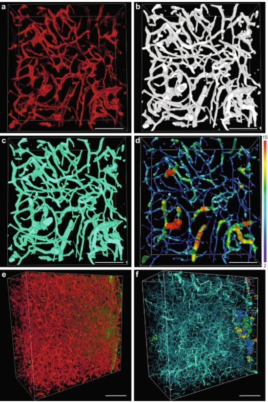
Precise methods for quantifying drug accumulation in brain tissue are currently very limited, challenging the development of new therapeutics for brain disorders. Transcardial perfusion is instrumental for removing the intravascular fraction of an injected compound, thereby allowing for ex vivo assessment of extravasation into the brain. However, pathological remodeling of tissue microenvironment can affect the efficiency of transcardial perfusion, which has been largely overlooked.
We... Read more
Serhii Kostrikov, Kasper B. Johnsen, Thomas H. Braunstein, Johann M. Gudbergsson, Frederikke P. Fliedner, Elisabeth A. A. Obara, Petra Hamerlik, Anders E. Hansen, Andreas Kjaer, Casper Hempel & Thomas L. Andresen
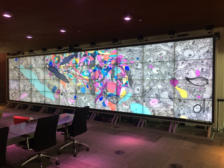
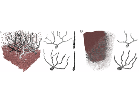
Insights into data with the KAUST Visualization Core Lab
Through collaboration, the KAUST Visualization Core Lab (KVL) team augments the efforts and domain expertise of KAUST researchers by providing complimentary technical knowledge with exploratory visualization and analytic tools.
KVL’s multi-year collaboration with KAUST Distinguished Professor P. Magistretti and research scientist C. Calì’s KAUST-EPFL Alliance for Integrative Modelling of Brain Energy Metabolism project—itself a collaboration with the Swiss Blue Br... Read more
By the KAUST Visualization Core Lab team and Caitlin Clark
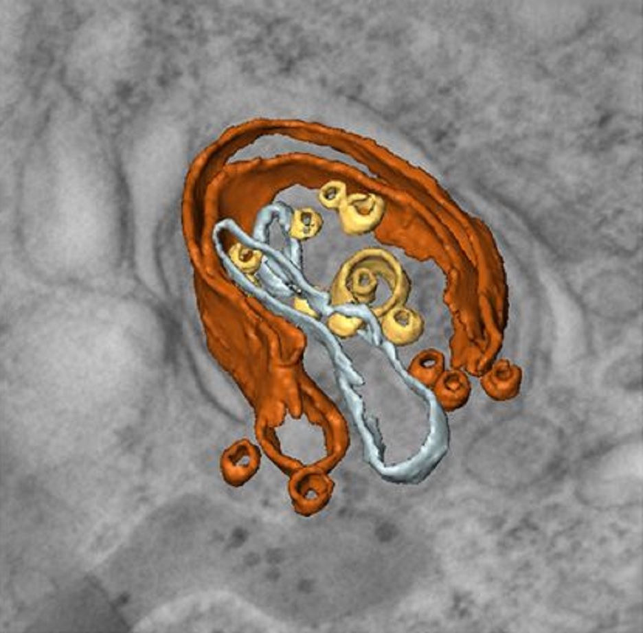
Macroautophagy is morphologically characterized by autophagosome formation. Autophagosomes are double-membraned vesicles that sequester cytoplasmic components for further degradation in the lysosome. Basal autophagy is paramount for intracellular quality control in post-mitotic cells but, surprisingly, the number of autophagosomes in post-mitotic neurons is very low, suggesting that alternative degradative structures could exist in neurons…
Read more
Maria Rosario Fernandez-Fernandez, Desire Ruiz-Garcia, Eva Martin-Solana, Francisco Javier Chichon, Jose L. Carrascosa, Jose-Jesus Fernandez
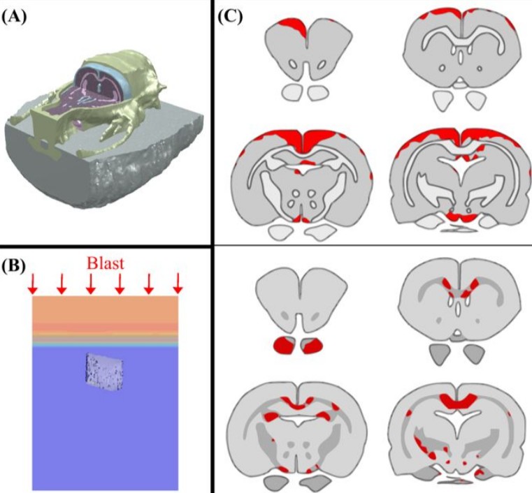
Cognition based bTBI mechanistic criteria; a tool for preventive and therapeutic innovations
Blast-induced traumatic brain injury has been associated with neurodegenerative and neuropsychiatric disorders. To date, although damage due to oxidative stress appears to be important, the specific mechanistic causes of such disorders remain elusive. Here, to determine the mechanical variables governing the tissue damage eventually cascading into cognitive deficits, we performed a study on the mechanics of rat brain under blast conditions. To this end, experiments were carried out to analyse... Read more
Daniel Garcia-Gonzalez, Nicholas S. Race, Natalie L. Voets, Damian R. Jenkins, Stamatios N. Sotiropoulos, Glen Acosta, Marcela Cruz-Haces, Jonathan Tang, Riyi Shi & Antoine Jérusalem
Three-dimensional virtual histology of human cerebellum by X-ray phase-contrast tomography
To quantitatively evaluate brain tissue and its corresponding function, knowledge of the 3D cellular distribution is essential. The gold standard to obtain this information is histology, a destructive and labor-intensive technique where the specimen is sliced and examined under a light microscope, providing 3D information at nonisotropic resolution. To overcome the limitations of conventional histology, we use phase-contrast X-ray tomography with optimized optics, reconstruction, and image an... Read more
Mareike Töpperwien, Franziska van der Meer, Christine Stadelmann, and Tim Salditt